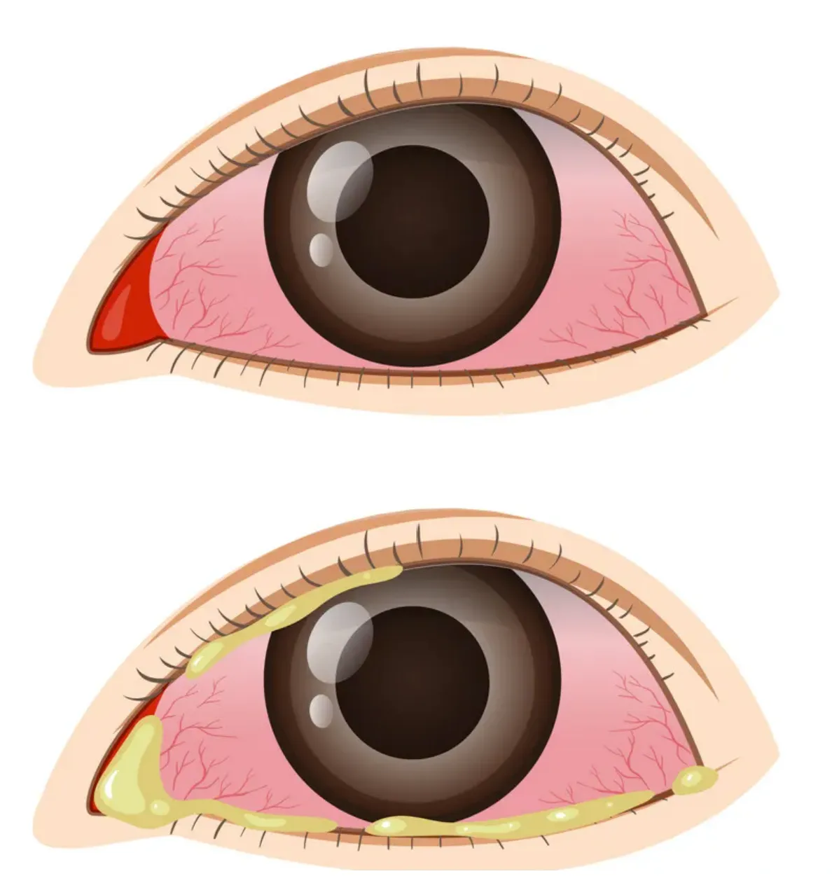
Conjunctivitis
Also called: Pink eye
Conjunctivitis, commonly known as “pink eye,” is an infection or swelling in your conjunctiva — the thin, transparent membrane that lies over the inner surface of the eyelid and covers the white part of your eye.
What your doctor is looking for during your eye exam: a plain-language tour of how the eye is put together, and the common eye conditions we check for at our Coconut Creek, Boynton Beach, Boca Raton and West Palm Beach offices.
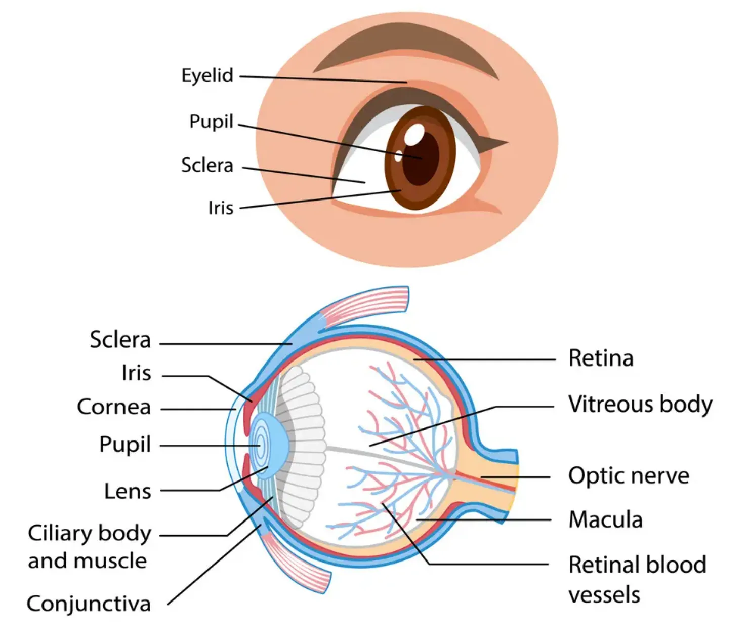
Your eye is a slightly asymmetrical globe, about an inch in diameter. The front part — what you see in the mirror — includes:
Just behind the iris and pupil lies the lens, which helps focus light on the back of your eye. Most of the eye is filled with a clear gel called the vitreous. Light projects through your pupil and lens to the back of the eye.
The inside lining of the eye is covered by special light-sensing cells collectively called the retina, which converts light into electrical impulses. Behind the eye, your optic nerve carries those impulses to the brain. The macula is a small, extra-sensitive area in the retina that gives you central vision — the detail and color you use when you look straight at something.
Want a second labeled illustration? See WebMD's picture of the eyes .
Six conditions we are often asked about — several of them far more common in children than parents expect. Each one is something a comprehensive eye exam is designed to find.
Also called: Pink eye
Conjunctivitis, commonly known as “pink eye,” is an infection or swelling in your conjunctiva — the thin, transparent membrane that lies over the inner surface of the eyelid and covers the white part of your eye.
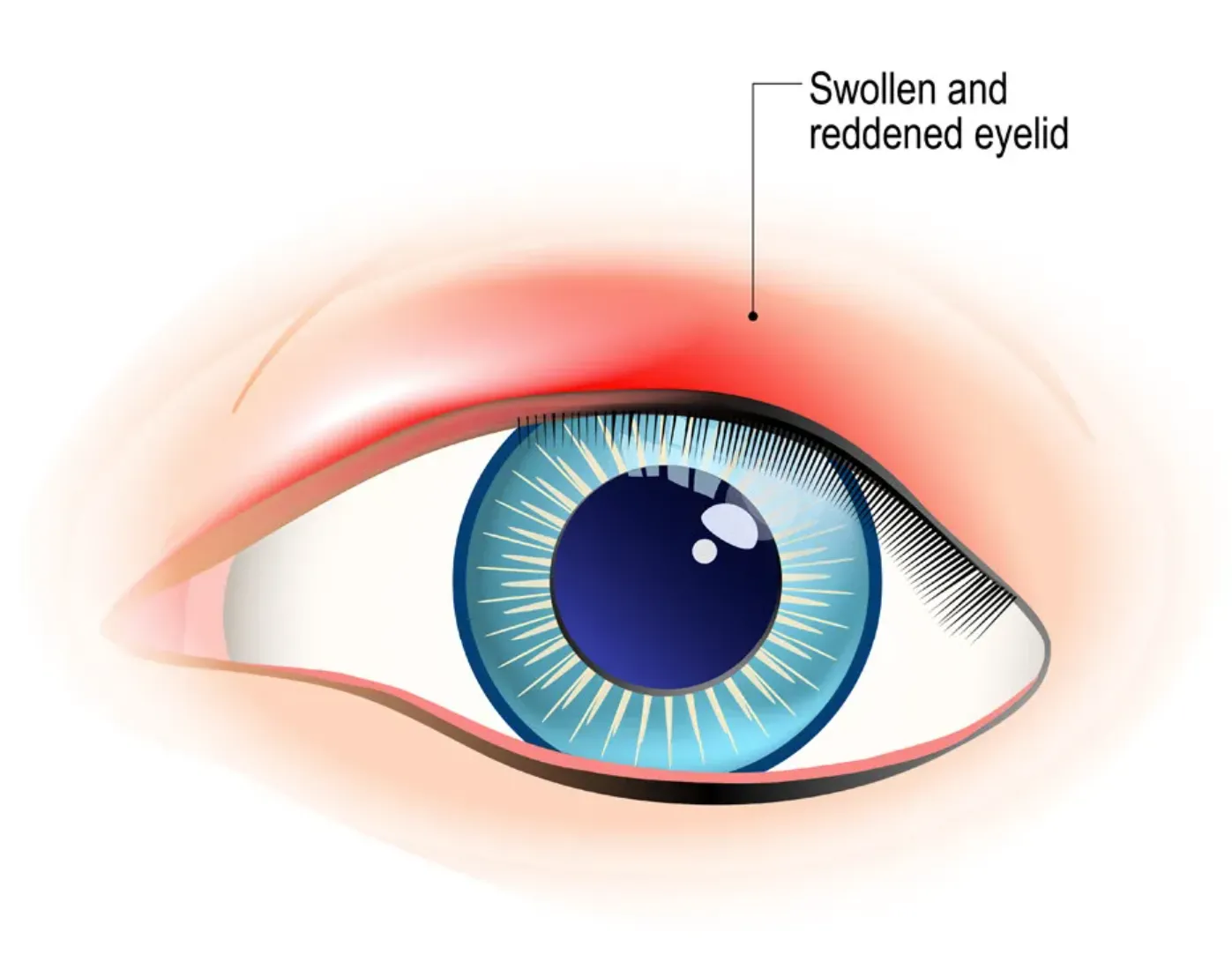
Blepharitis is inflammation of the eyelid margin. It is commonly a chronic condition that affects both children and adults. It is generally benign, but it can cause frequent and annoying symptoms.
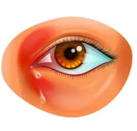
Dacryocystitis is an infection of the tear (lacrimal) sac, usually due to a blockage in the tear (nasolacrimal) duct.
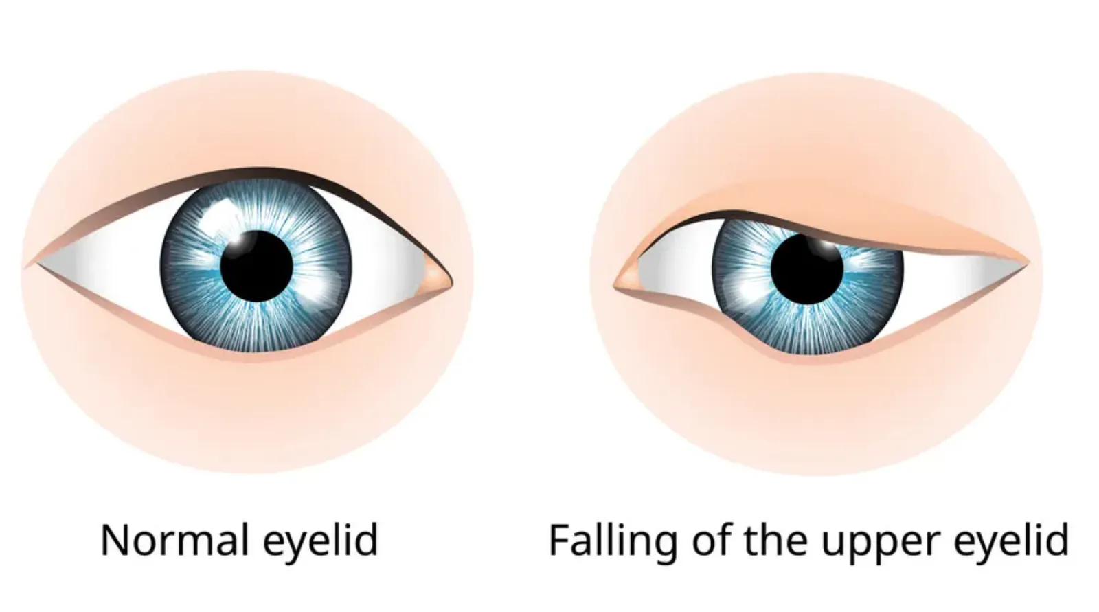
Also called: Droopy eyelid
A droopy eyelid, also called ptosis, occurs when the muscle that elevates the eyelid — the levator palpebrae superioris — is weak, for a number of possible reasons.
The most common cause in children is a levator palpebrae superioris that did not develop well. That form is present at birth and is called congenital ptosis.
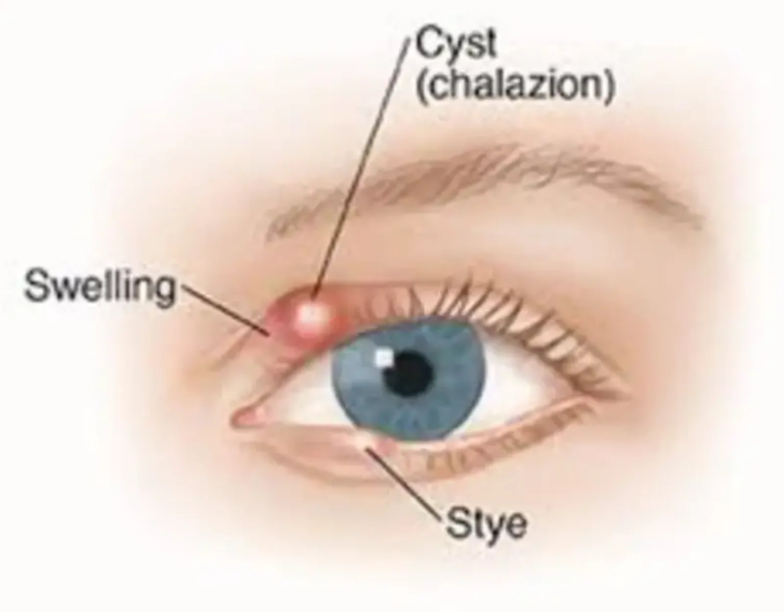
A chalazion is a localized bump in the eyelid, of varying size. Small glands lining the edge of the eyelids (the meibomian glands) produce oil that helps lubricate the surface of the eye. When one of those glands becomes blocked, oil backs up inside the gland and forms a bump in the eyelid.
Breakdown of the oily secretions causes inflammatory cells to rush to the area. That irritates the surrounding eyelid skin, causing it to become red and swollen, and sometimes painful.
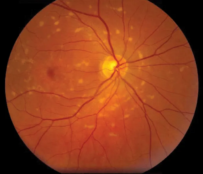
Also called: Juvenile macular degeneration
Stargardt disease is an inherited form of macular degeneration that affects children and young adults. It causes deterioration of the cells in the center of the retina that convey information to the brain.
Over time, Stargardt disease causes a blurred, distorted, dark, or empty area in the center of vision. The center of the retina — the macula — is responsible for our ability to see color and fine detail when we look directly at an object, so diseases like Stargardt affect a person’s ability to read and to drive.
These descriptions are educational, not a diagnosis. A red, swollen or painful eye, a lid that has started to droop, or any sudden change in vision should be examined promptly. Contact us or book an appointment and we will take a look, explain what we find, and coordinate care or a referral when that is what the situation calls for.
Every comprehensive evaluation at Vision & Learning Center includes an assessment of overall eye health — the front of the eye and the structures behind it, the lids and tear system, and the retina, macula and optic nerve at the back. Finding and managing an eye-health concern early is how long-term sight is protected. When we find something medical, we explain it in plain language and coordinate care or a referral as appropriate.
Healthy eyes and sharp eyesight are the foundation — but they are not the whole picture. Eyes can be perfectly healthy and still not work well together, and that is where reading fatigue, headaches, losing your place on a page and double vision come from. Our exams check both: the health of the eye, and the visual skills — eye teaming, focusing and tracking — that a school screening never measures. Learn more about our examinations, or start with the symptom checklist if something already feels off.
Screens, short-sightedness and eye health, separating what is established from what is repeated.
Whether it is a red eye, a lid that looks different, or a child who is struggling to read, an exam at our Coconut Creek, Boynton Beach, Boca Raton or West Palm Beach office tells you what is actually going on.