Learn More About

Cranial Nerve Palsy

Cranial nerve palsies involve impairment of the cranial nerves, often affecting motor or sensory functions — including vision and eye movement. When the nerve involved is one of the three that aim the eyes, the eyes stop pointing at the same place and double vision follows.

Third, fourth & sixth nerves Children & adults 4 South Florida offices
Start Here

What a Cranial Nerve Palsy Is

A cranial nerve palsy is an impairment of one of the cranial nerves, and it often affects motor or sensory function — including vision and eye movement. Three of those nerves carry the signals that aim the eyes: the oculomotor nerve (CN III), the trochlear nerve (CN IV) and the abducens nerve (CN VI).

When one of them is impaired, the muscle it supplies no longer pulls its share. The affected eye drifts out of alignment with its partner, the brain receives two images it cannot merge, and the result is double vision (diplopia) — often with a compensating head tilt or turn, a drooping lid, or the habit of closing one eye for relief. Which nerve is involved determines which direction the eye drifts and which movements become difficult.

Sudden, new or worsening double vision, a newly drooping eyelid or a newly dilated pupil should be evaluated promptly by a physician. This page is educational and is not a diagnosis.

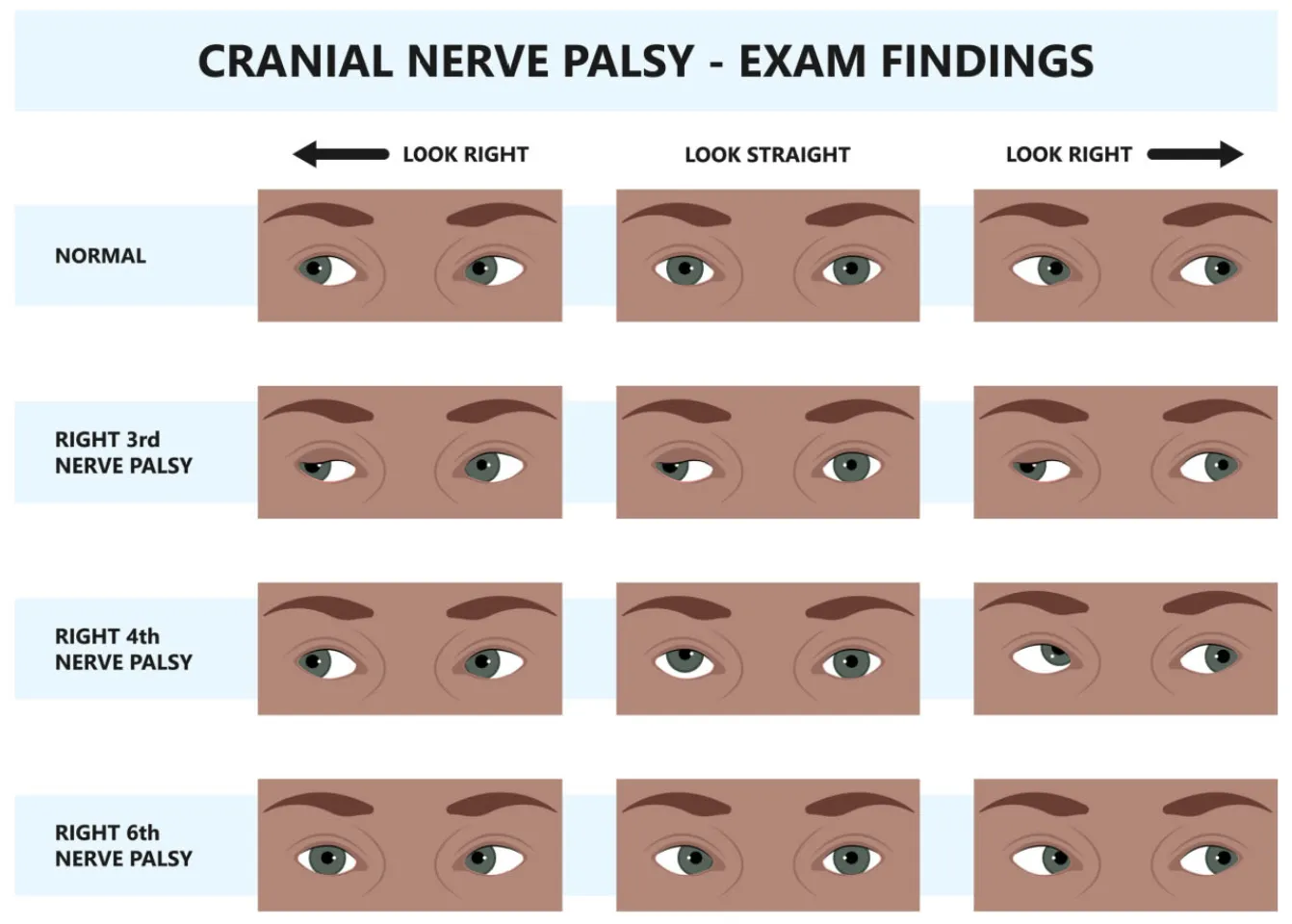
Chart of cranial nerve palsy exam findings, comparing normal eye movements with right third, fourth and sixth nerve palsies on left gaze, straight-ahead gaze and right gaze.
How each palsy shows up on examination: the eyes are compared on left gaze, straight ahead and right gaze.
The Three Patterns

The Nerves That Move the Eyes

Muscles affected, symptoms and common causes for each of the three eye-movement nerves.

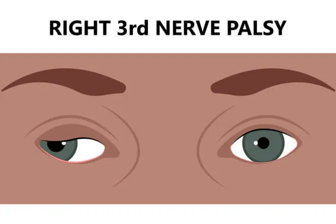
Illustration of a right third nerve palsy: the right upper eyelid droops over a pupil that sits low and turned outward.

Oculomotor Nerve Palsy

(Cranial Nerve III)

Muscles affected
Most eye muscles, the levator palpebrae (the eyelid muscle), and the muscle responsible for pupil constriction.
Symptoms
Drooping eyelid (ptosis), dilated pupil, eye turned down and out, double vision.
Causes
Ischemia (due to diabetes or hypertension), aneurysm, tumor, trauma, or inflammation.
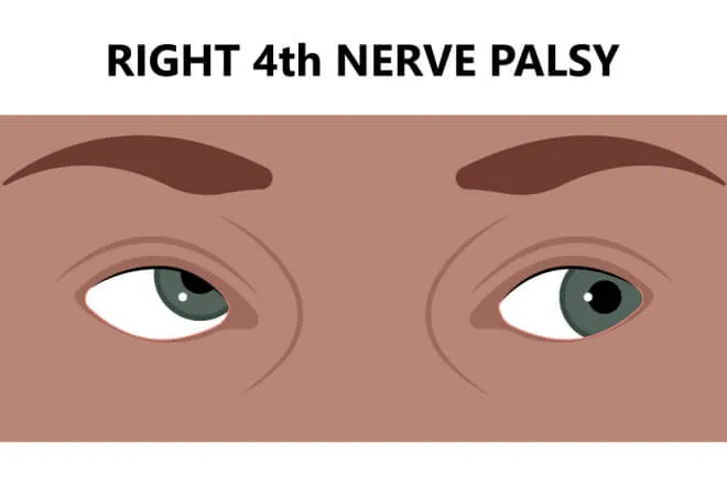
Illustration of a right fourth nerve palsy: the right eye sits higher than the left when looking straight ahead.

Trochlear Nerve Palsy

(Cranial Nerve IV)

Muscles affected
Superior oblique muscle.
Symptoms
Head tilt, difficulty looking down — especially when the eye is turned inward — and double vision.
Causes
Head trauma, congenital defects, tumor, or ischemia.
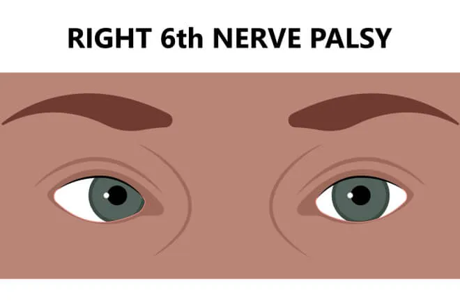
Illustration of a right sixth nerve palsy: the right eye stays turned inward and cannot rotate outward.

Abducens Nerve Palsy

(Cranial Nerve VI)

Muscles affected
Lateral rectus muscle.
Symptoms
Inability to abduct the eye (turn it outward), double vision.
Causes
Increased intracranial pressure, ischemia, tumor, or inflammation.
Management

Managing the Visual Symptoms

Three tools are used, alone or together, to make seeing single and comfortable again while the underlying cause is treated.

Vision Therapy

Vision therapy, also known as orthoptic therapy, can be part of the management strategy for cranial nerve palsies — especially when they cause ocular misalignment and binocular vision dysfunction such as double vision (diplopia). It works on eye coordination, focusing ability and more efficient visual processing.

  • Specific eye exercises can help improve muscle strength and coordination, which may help reduce symptoms like double vision.
  • Techniques that train the brain to fuse the images from each eye can help in managing double vision.
  • Biofeedback and other visual feedback methods can help refine visual skills and improve visual performance.
  • Computer-based games can aid in developing sensory fusion.

Prism Glasses

Prism bends light before it reaches the eye, so the two images the brain receives line up more comfortably.

  • Prism glasses can be beneficial in managing double vision by aligning the two images into one.
  • They can be used temporarily or permanently, depending on the progress and response to therapy.

Occlusion Therapy

When two images cannot yet be fused, removing one of them removes the confusion while the underlying cause is treated.

  • Temporarily occluding one eye with a patch or a fogged lens can alleviate double vision, and can be helpful especially in the initial stages.
Honest Expectations

A Comprehensive Approach

It is important to understand that vision therapy is not a cure for cranial nerve palsy, but it can be a valuable tool in managing the symptoms and improving visual function. The suitability and success of vision therapy depend on the individual case, including the type and degree of the cranial nerve palsy and the patient's age, compliance and overall health.

Addressing the underlying cause of the palsy is equally crucial. Managing diabetes, reducing intracranial pressure or treating other underlying conditions matters for overall management, and in some cases surgical intervention may be required to realign the eyes or address the cause of the palsy itself.

Vision care is one part of the plan. We work on the visual consequences of a palsy — alignment, fusion and comfort — while your medical team treats the cause. Care is coordinated, not competing.
Follow-Up

Regular Monitoring

Cranial nerve palsies change over time — some recover, some stabilize, some need the plan adjusted along the way. Regular monitoring and follow-up with an optometrist or ophthalmologist specializing in binocular vision and neuro-optometry are crucial to assess progress and modify the treatment plan as needed.

A neurologist should also be involved in the overall management and treatment of the palsy, to address the neurological aspects and the underlying causes. Where a head injury is part of the picture, neuro-optometric rehabilitation is often coordinated with physical and occupational therapy.

A comprehensive developmental and binocular vision evaluation is where the visual side of the plan starts: how the eyes align and team at distance and near, how they track and change focus, and how the whole system holds up under sustained work. Children and adults are seen at our Coconut Creek, Boynton Beach, Boca Raton and West Palm Beach offices.
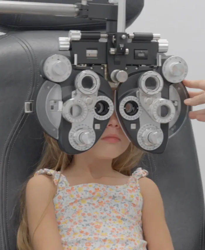
A child looking through a phoropter during an eye examination
Questions

Frequently Asked Questions

Can vision therapy cure a cranial nerve palsy?

No. Vision therapy is not a cure for cranial nerve palsy, but it can be a valuable tool in managing the symptoms and improving visual function — particularly the ocular misalignment and double vision that come with it. Whether it is suitable, and how much it helps, depends on the individual case: the type and degree of the palsy, and the patient’s age, compliance and overall health.

Which cranial nerves affect the eyes?

Three of them carry the signals that aim the eyes. The oculomotor nerve (CN III) supplies most of the eye muscles, the eyelid muscle and pupil constriction; the trochlear nerve (CN IV) supplies the superior oblique; and the abducens nerve (CN VI) supplies the lateral rectus. Which nerve is involved shapes the pattern of misalignment and the symptoms — see the three patterns.

What else needs to be treated besides the eyes?

Addressing the underlying cause is crucial. Managing diabetes, reducing intracranial pressure or addressing other underlying conditions matters for overall management, and in some cases surgical intervention may be required to realign the eyes or address the cause of the palsy. A neurologist should be involved in the overall management and treatment to address the neurological aspects and underlying causes.

Do prism glasses have to be permanent?

Not necessarily. Prism can be used temporarily or permanently, depending on progress and the response to therapy. In the initial stages, temporarily occluding one eye with a patch or a fogged lens is another way to relieve double vision while the underlying cause is addressed.

Who should follow a cranial nerve palsy over time?

Regular monitoring and follow-up with an optometrist or ophthalmologist who specializes in binocular vision and neuro-optometry are crucial to assess progress and modify the treatment plan as needed, alongside the neurologist managing the underlying cause. We see patients at {evaluationOfficePhrase()}.

Next Steps

Start With an Evaluation

If double vision, a head tilt or a drooping lid is part of your daily life, an evaluation measures exactly how the two eyes are working together — and what can be done to make seeing single and comfortable again.