Strabismus Surgery and Vision Therapy: Why Combine Them
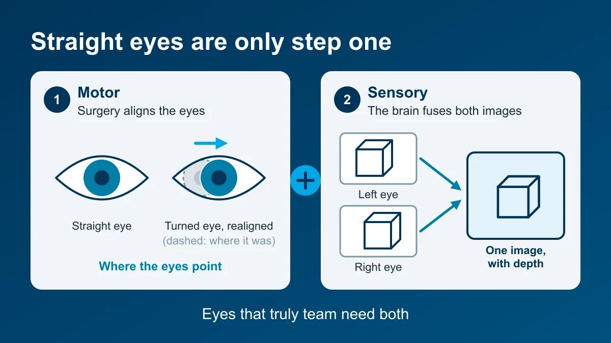
Strabismus surgery and vision therapy are often presented as rival choices: operate on the eye muscles, or train the eyes. A 2025 review written jointly by a developmental optometrist and a pediatric strabismus surgeon makes a stronger case: for many people, the best results come from using the two together (Del Campo & Lenart, 2025). Surgery puts the eyes in the right place. Vision therapy trains the brain to use them as a pair, which can help the result hold.
The controlled evidence points the same way. In a randomized trial of children with intermittent exotropia, adding therapy after a successful operation cut the rate of suboptimal outcomes at a year roughly in half, from 42.6% to 20.5%, with better depth perception and eye-teaming control (Xu et al., 2023).
Here is how the combined approach works: why straight eyes after surgery don’t automatically work together, where therapy fits before and after an operation, what the authors’ five cases achieved, and what the trials show. It is written for parents weighing surgery for a child and for adults whose eyes were straightened years ago but still don’t work as a pair.
Key takeaways
- Surgery changes where the eyes point. Whether the brain then combines the two images into one, with depth, is a separate sensory skill, and that skill can be trained. That is the core case for pairing surgery and therapy.
- In a randomized trial of 136 children with intermittent exotropia, therapy after surgery halved suboptimal outcomes at 12 months, 20.5% versus 42.6%, and improved stereopsis and fusional control (Xu et al., 2023).
- A 2025 review by a developmental optometrist and a strabismus surgeon describes three roles for vision therapy: building visual skills, “sensory readiness” before surgery, and a “binocular safeguard” after it (Del Campo & Lenart, 2025). Their five patients, aged 12 to 53, all improved, and three who had no measurable stereopsis gained it.
- The same authors suggest surgery be discussed when an esotropia exceeds 15 to 20 prism diopters, an exotropia 20 to 25, or a vertical turn 10.
- Alignment alone often slips. In PEDIG’s childhood exotropia trial, 11% to 30% of children, depending on the operation, needed a second surgery by eight years (Donahue et al., 2024). In a trial of children with intermittent exotropia, building fusion after surgery reduced suboptimal outcomes at 12 months; longer-term effects haven’t been studied.
Why straight eyes are not the whole goal
Strabismus is common. In insurance claims data on more than 10 million US children, it was the most frequently diagnosed significant eye condition, at 3.2% (Pineles et al., 2022). Adults are not exempt: the lifetime risk of a new eye turn in adulthood is about 4% (Martinez-Thompson et al., 2014). Our post on adult strabismus and double vision covers those adult causes and red flags.
The effects reach beyond appearance. Adults with amblyopia and strabismus describe trouble judging distances when driving, reading for long periods, catching a ball and using stairs (Kumaran et al., 2019). In a small study, children with infantile strabismus scored lower on a motor-coordination questionnaire than matched classmates, and those without stereopsis scored lowest (Vagge et al., 2021). Children with amblyopia also tend to read slowly, which is why researchers recommend classroom accommodations such as extra time (Birch & Kelly, 2017).
What the brain does when the eyes don’t line up
When the eyes point at different places, the brain receives two conflicting pictures. A developing visual system protects itself in a few ways, and surgery alone does not undo these adaptations (Del Campo & Lenart, 2025):
- Suppression. The brain switches off part of the turned eye’s input to avoid double vision. It is a sensible shortcut that becomes a habit.
- Amblyopia. Reduced best-corrected sight in one eye, often alongside the suppression.
- Anomalous correspondence. Put simply, the brain redraws its map, so the turned eye’s “straight ahead” point is no longer its center. The brain has learned to make a crooked pair of eyes feel matched. The catch is that when a surgeon straightens the eyes, the old map may now feel wrong, which can bring double vision or a pull back toward the old position.
- Weak eye-movement and focusing skills. The authors describe frequent problems with smooth tracking, quick eye jumps, focusing and eye-teaming ranges in this group.
Real teamwork has levels: seeing with both eyes at once, fusing the two images, and then stereopsis, the fine depth sense. Our explainer on the three grades of binocular vision goes through them, and our guide to how depth perception develops explains what strabismus does to it.
Does surgery alone restore depth perception?
Sometimes, but not reliably. In 24 adults with long-standing, early-onset strabismus and no fusion before surgery, all showed peripheral fusion after being aligned, and half reached stereoacuity of 200 seconds of arc or better (Morris et al., 1993). Among 132 adults operated on for any cause, 48% ended up with stereoacuity better than 100 seconds of arc and 21.5% had none; people whose strabismus began in childhood were least likely to regain it (Rock et al., 2025).
A study of 73 patients with childhood-onset strabismus, all aligned within 10 prism diopters, found poorer stereopsis with amblyopia, esotropia, onset before six months and a larger turn before surgery. Only one patient in the study gained binocular single vision after surgery (Singh et al., 2022). Straight eyes open the door. They don’t guarantee anyone walks through it.
The treatment options for strabismus
Every plan starts with ruling out disease, a careful history and a full binocular workup, then the right glasses. That may include a reading add for accommodative esotropia, prism, or partial occlusion. If the turn or symptoms persist, the authors lay out four paths (Del Campo & Lenart, 2025):
| Option | What it involves | Where the authors see it fitting |
|---|---|---|
| Vision therapy alone | Weekly in-office sessions and home practice to build fusion, eye movements and focusing | Smaller or intermittent turns and skill deficits; they note it may not be enough for large angles |
| Surgery alone | A surgeon tightens or loosens eye muscles to reduce the turn | Addresses alignment, but by itself not fusion, stereopsis or sensory adaptations |
| Combined | Therapy before and/or after surgery, with the optometrist and surgeon sharing findings | Their preferred route for larger turns, especially with childhood onset, amblyopia or prior surgery |
| No treatment | Monitoring | Discussed openly, including the risk of continued symptoms or progression |
When should strabismus surgery be discussed?
The authors suggest surgery belongs in the conversation when the turn is large or frequent and symptomatic, and the person is a suitable candidate. Their size thresholds are an esotropia (inward turn) greater than 15 to 20 prism diopters, an exotropia (outward turn) greater than 20 to 25, or a vertical turn greater than 10. They note that surgical success is usually defined in the literature as a remaining misalignment of less than 10 prism diopters at distance and near.
That definition is about position, and the authors’ point is that it leaves out fusion, stereopsis and how well the person actually sees. Appearance is a legitimate reason to consider treatment, too; our post on the social side of eye turns and patching looks at what that research measured. If a child reports seeing double, start with double vision in children.
Vision therapy before and after strabismus surgery: three roles
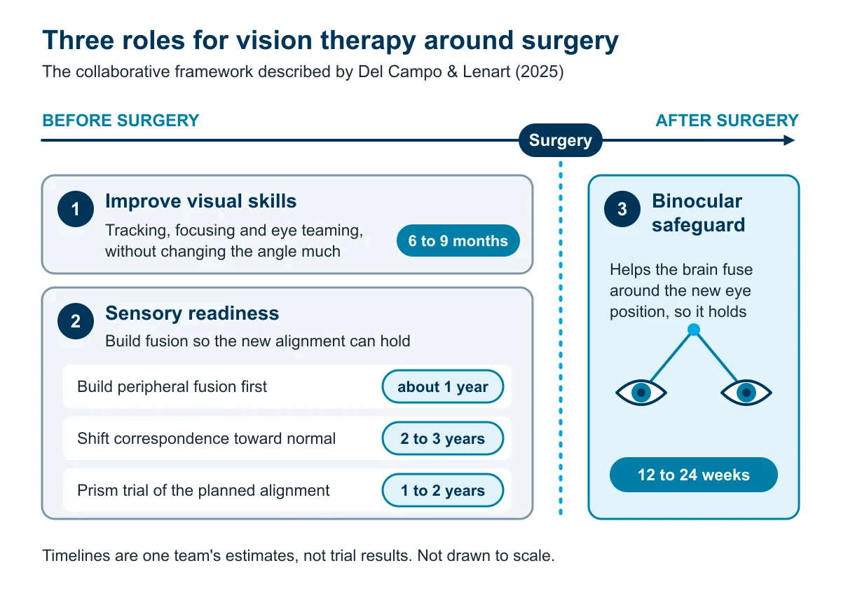
The heart of the review is three distinct jobs for therapy, each with its own timeline (Del Campo & Lenart, 2025):
1. Improving visual skills. Eye tracking, focusing and eye-teaming problems are treated without deliberately disturbing the sensory adaptation or changing the angle much. The authors expect this to take 6 to 9 months. For people with childhood-onset strabismus whose main goal is appearance, they recommend it before surgery.
2. Sensory readiness before surgery. The aim is to build the brain’s ability to combine the two eyes’ images, so that once the surgeon straightens them there is fusion to hold them there. The authors describe three versions:
- Building fusion in the peripheral vision and working around a residual anomalous map or central suppression: about 1 year.
- Going further, trying to shift anomalous correspondence toward normal with progressively finer targets: about 2 to 3 years, and not everyone responds.
- Trialing prism to preview the planned alignment, which also helps the surgeon choose the amount of correction: about 1 to 2 years, variable.
3. A binocular safeguard after surgery. Therapy helps the brain learn to fuse around the new eye position, which makes it more likely to hold. This is the role with randomized-trial support (see below). The authors estimate 12 to 24 weeks, depending on evaluation findings.
One line from the review is worth underlining: a patient who fails to develop any binocular cooperation through therapy is also likely to be a poor surgical candidate. In the authors’ model, pre-surgical therapy aims to build what the visual system can do and shows the surgical team what to expect before the operation.
How the optometrist and surgeon share the work
In the authors’ model, the optometrist runs the expanded sensory testing and may recommend how many prism diopters they think the surgery should correct, based on how the patient fuses with prism. The surgeon makes the final decision on the muscle adjustment. The therapist’s observations, such as when reliable peripheral fusion appears, help time the referral.
Surgeons differ on targets, too. The authors describe deliberately under-correcting exotropia at first to avoid overshooting into an inward turn. A 2026 PEDIG analysis of children found the opposite association: a small inward overshoot one week after exotropia surgery went with fewer recurrences over eight years, at the cost of slightly more problematic esotropia (PEDIG, 2026). This is a question for your surgeon.
The five case examples in the article
The review illustrates the framework with five cases treated by the authors’ collaborating teams. The results are striking: patients aged 12 to 53, several of them adults whose earlier surgery had not held, reached single vision or straighter eyes, and the three who had no measurable stereopsis gained it. They are the authors’ patients, not ours, and they are summarized here from the article.
| Case | Age and history | What was done | Reported outcome |
|---|---|---|---|
| 1 | 44, childhood esotropia, operated at 15; relying on more prism, double vision at distance | Weekly therapy for about two years, surgery partway through, planned with prism data from therapy | Single vision at all distances without prism; stable and symptom-free at 1.5 years. Stereo was already present and changed little |
| 2 | 51, childhood exotropia that became constant | First surgery, then weekly therapy, then a second surgery | A large constant outward turn became a small turn showing less than 10% of the time; stereo moved from coarse to moderate, with some sense of depth on a random-dot test |
| 3 | 31, eyes straight after surgery, then the old alternating outward turn returned | 12 weeks of therapy, started more than a year after surgery, with no further operation | Eyes straight on cover testing at a 2-month follow-up; stereo went from none to fine local stereo |
| 4 | 53, childhood esotropia, operated at 12 with no gain in depth and a return of the turn; anomalous correspondence | Therapy before and after a second surgery | Turn reduced to small and occasional; stereo went from none to coarse; reported everyday 3D viewing |
| 5 | 12, constant esotropia with anomalous correspondence and mixed amblyopia | About two years of therapy, surgery near the end | Weaker eye improved from 20/40 to about 20/20; stereo from none to moderate; parents reported more confidence and stamina, and he reported seeing 3D |
Case 3 stands out: an adult whose turn had returned after surgery was straight again after 12 weeks of therapy, with no second operation. As case reports without a comparison group, these illustrate the approach rather than prove it, and the authors call for larger studies.
What the controlled evidence shows
The “safeguard” idea has been put to a randomized test, and it held up. A trial in China enrolled 136 children aged 7 to 17 whose intermittent exotropia was well aligned a month after surgery (Xu et al., 2023). Half had at least 8 weeks of hospital-based therapy, one or two sessions a week, plus home practice; half had routine follow-up. By 12 months, 20.5% of the therapy group had a suboptimal result versus 42.6% of controls: therapy roughly halved the rate. Stereopsis and fusional control were better with therapy too.
The benefit came even though fewer than half of the therapy group completed the full program. The trial was single-center with no sham therapy, and it studied school-age children with intermittent exotropia, so results in infantile esotropia and adults rest on clinical reports like the cases above.
More broadly, a 2024 major review of intermittent exotropia found that surgery reduces the angle and improves stereopsis and quality of life, but with a substantial tendency toward reoperation or recurrence, and that no randomized trial has compared surgery with observation (Ma & Scheiman, 2024).
Does strabismus come back after surgery?
It can. In PEDIG’s randomized trial of 197 children with intermittent exotropia, 37% to 46% had a suboptimal result by three years, depending on the operation (PEDIG, 2019). By eight years, reoperation had occurred in 30% after operating on both outer muscles and 11% after a recess-resect procedure (Donahue et al., 2024). In adults, the American Academy of Ophthalmology’s review reported successful realignment in 68% to 85% of large case series, unplanned reoperation in up to 21% of comitant cases, and new double vision after surgery in 1% to 14% (Mills et al., 2004).
Those numbers are the strongest argument for the collaborative model. In the review authors’ view, weak fusion and an unaddressed anomalous map help explain why alignment drifts back, and building fusion around the new position helps it hold. The randomized trial above found just that pattern: fewer suboptimal outcomes and better fusional control with therapy.
Questions to ask if you or your child is considering strabismus surgery
The review’s shared-decision list covers five things: the diagnosis, the performance challenges it causes at school, work and in daily life, your goals, the nature and purpose of the proposed treatment, and the likely outcome of each option, including no treatment. Turned into questions:
- What exactly is the diagnosis? Which direction is the turn, how large is it at distance and near, and is it constant or intermittent?
- How is it affecting daily life? Reading, sports, driving, eye contact, double vision or eye strain.
- What are our goals? Appearance, single vision, depth perception, comfort, or all of these. Say them out loud, and write them down.
- What does the surgery aim to do, and how is success defined? Ask whether the target is alignment alone or also fusion and stereopsis.
- What are the chances of each outcome? Including a second operation, and what happens if we wait.
- Has sensory testing been done? Suppression, fusion, anomalous correspondence and stereopsis, not just the angle.
- Would therapy before surgery improve the odds, and would therapy afterward help it hold?
- How will the surgeon and the therapy provider share findings? Including prism measurements before surgery.
- What follow-up is planned, and for how long?
If amblyopia is part of the picture, our summary of amblyopia treatment options and the PEDIG trials explains glasses, patching and drops.
Our clinical perspective
We are a developmental optometry practice, and this collaborative model is how we work. We do not perform eye muscle surgery. When a turn is large and constant, we co-manage with the surgeon, and our role looks a lot like the one this review describes: measuring the sensory side carefully, building fusion before surgery where it is weak, sharing prism and fusion findings, and helping the brain use the new alignment afterward.
In our practice we regularly see what that brings: eyes that stay straighter, double vision that settles, reading that becomes more comfortable, and depth perception that people notice in daily life, from catching a ball to watching a 3D movie. Families often tell us their child seems more confident once the eyes work as a team. For smaller and intermittent turns, therapy on its own is often enough. Our graduates Amelia and Alex both had eye turns and no depth perception before vision therapy, and in each family’s experience gained it with therapy alone; more patients describe their results on our success stories page.
Every visual system is different, so outcomes vary and a careful evaluation comes first. The physiology, the case reports and a randomized trial in children all point the same way: straight eyes are the start, and teaching them to work together helps the result hold.
If you or your child has an eye turn, has had surgery and still struggles with depth, double vision or reading comfort, or is weighing surgery now, start with a full binocular evaluation. See what our amblyopia and strabismus care and vision therapy involve, read about lazy eye and our examinations, find a location near you, or schedule an evaluation.
Common Questions About This Topic
Do you need vision therapy after strabismus surgery?
It can make a real difference. In a randomized trial of 136 children with intermittent exotropia, therapy after surgery halved the rate of suboptimal outcomes at 12 months, 20.5% versus 42.6%, with better stereopsis and fusional control. It matters most for people whose eyes are straighter but not yet working as a pair, or whose alignment has a history of slipping. Some people fuse and gain depth perception from surgery alone, which a full binocular evaluation helps identify.
Can vision therapy replace strabismus surgery?
For small or intermittent turns and eye-teaming problems, therapy on its own is often the right treatment. For a large, constant turn, many clinicians, including the review's authors, recommend using the two together. A 2025 review by a developmental optometrist and a strabismus surgeon suggests discussing surgery when an esotropia is over 15 to 20 prism diopters, an exotropia over 20 to 25, or a vertical turn over 10, with therapy used alongside the operation rather than instead of it.
Does strabismus come back after surgery?
It can. In PEDIG's randomized trial of childhood intermittent exotropia surgery, 30% of children who had both outer muscles recessed and 11% who had a recess-resect procedure needed a second operation by eight years. The American Academy of Ophthalmology's review of adult surgery reported unplanned reoperation in up to 21% of comitant strabismus cases. That is why follow-up matters, and why, in a trial of children with intermittent exotropia, therapy after surgery was used to help the new alignment hold.
Will my child have depth perception after strabismus surgery?
Straight eyes make depth perception possible but do not guarantee it. In a study of 73 patients with childhood-onset strabismus aligned within 10 prism diopters, amblyopia, esotropia, onset before six months of age and a larger turn before surgery were all linked to poorer stereopsis afterward. Only one patient in that study gained binocular single vision after surgery, which is why some teams, including the review authors, work on fusion alongside surgery.
How long is vision therapy before or after strabismus surgery?
It depends on the goal. In the 2025 collaborative framework, therapy for visual skills alone is expected to take about 6 to 9 months. Building fusion before surgery takes about 1 year, or 2 to 3 years when the aim is to change anomalous correspondence. Trialing prism before surgery takes about 1 to 2 years. The post-surgery safeguard phase runs about 12 to 24 weeks. These are the authors' clinical estimates.
Where This Information Comes From
- Del Campo S, Lenart T (2025). Collaborative Management of Strabismus. Advances in Ophthalmology and Optometry; 10:17-36. : Narrative review by a developmental optometrist and a strabismus surgeon who work together, with five illustrative cases (ages 12 to 53) from their collaborating clinics. Describes three roles for vision therapy around surgery, suggested size thresholds for discussing surgery and timelines for each role. Not a controlled study; the authors call for case series, prospective studies and clinical trials. No PubMed record was found for this article.
- Pineles SL, Repka MX, Velez FG, et al. (2022). Prevalence of pediatric eye disease in the OptumLabs Data Warehouse. Ophthalmic Epidemiology; 29(5):537-544. : Claims data on 10,759,066 commercially insured US children, 2007-2018: strabismus was the most common significant eye diagnosis at 3.2%, followed by amblyopia at 1.5%. Claims-based, so it may reflect access to care as well as true prevalence.
- Martinez-Thompson JM, Diehl NN, Holmes JM, Mohney BG (2014). Incidence, types, and lifetime risk of adult-onset strabismus. Ophthalmology; 121(4):877-882. : Population-based cohort in Olmsted County, Minnesota: 753 new adult cases over 20 years; lifetime risk of adult-onset strabismus 4.0% in women and 3.9% in men; paralytic strabismus most common at 44.2%.
- Kumaran SE, Khadka J, Baker R, Pesudovs K (2019). Functional limitations recognised by adults with amblyopia and strabismus in daily life: a qualitative exploration. Ophthalmic and Physiological Optics; 39(3):131-140. : Interviews and focus groups with 37 adults (62% strabismic amblyopia): reported difficulties judging distances and changing lanes while driving, prolonged reading, catching a ball, stairs and work tasks. Qualitative, so it describes experiences rather than measuring how common they are.
- Vagge A, Pellegrini M, Iester M, et al. (2021). Motor skills in children affected by strabismus. Eye (London); 35(2):544-547. : 23 children aged 5-11 with infantile strabismus versus 24 matched controls: motor-coordination questionnaire scores were lower with strabismus (58.7 vs 74.2), and lowest in those without stereopsis. Small, parent-questionnaire outcome.
- Birch EE, Kelly KR (2017). Pediatric ophthalmology and childhood reading difficulties: Amblyopia and slow reading. Journal of AAPOS; 21(6):442-444. : Commentary summarizing research that children with amblyopia read slowly, and recommending school accommodations such as extra time. Opinion piece, not a new study.
- Singh A, Patnaik N, Mittal SK, et al. (2022). Factors Determining Improvement in Stereopsis and Binocularity After Good Postoperative Alignment in Patients With Childhood-Onset Strabismus. Cureus; 14(2):e21964. : Prospective series of 73 patients aged 5-35 with constant childhood-onset strabismus over 30 prism diopters, aligned within 10 after surgery: amblyopia, esotropia, onset before six months and a preoperative turn over 45 were linked to poorer stereopsis; only one patient gained binocular single vision postoperatively. Single center, 12-week follow-up.
- Rock O, Golani T, David S, et al. (2025). Improvement of stereopsis following strabismus surgery in adults: a retrospective analysis. Canadian Journal of Ophthalmology; 60(4):e598-e601. : 132 adults operated on for any cause, 2010-2022: after surgery 48% had stereoacuity better than 100 seconds of arc and 21.5% had none; postoperative angle was the strongest factor, and childhood strabismus had the lowest chance of regaining stereopsis. Retrospective, single institution.
- Morris RJ, Scott WE, Dickey CF (1993). Fusion after surgical alignment of longstanding strabismus in adults. Ophthalmology; 100(1):135-138. : 24 adults with early-onset strabismus and no fusion before surgery: after alignment within 8 prism diopters, all showed peripheral fusion and 12 (50%) reached 200 seconds of arc or better. Small, uncontrolled.
- Mills MD, Coats DK, Donahue SP, Wheeler DT; American Academy of Ophthalmology (2004). Strabismus surgery for adults: a report by the American Academy of Ophthalmology. Ophthalmology; 111(6):1255-1262. : Review of 49 reports: successful realignment in 68-85% of adults in large case series; new postoperative double vision 1-14%; unplanned reoperation up to 21% in comitant strabismus and up to 50% in thyroid eye disease. Mostly level III evidence.
- Pediatric Eye Disease Investigator Group; Donahue SP, Chandler DL, Holmes JM, et al. (2019). A Randomized Trial Comparing Bilateral Lateral Rectus Recession versus Unilateral Recess and Resect for Basic-Type Intermittent Exotropia. Ophthalmology; 126(2):305-317. : 197 children aged 3 to under 11: suboptimal surgical outcome by 3 years in 46% after bilateral recession versus 37% after recess-resect, not statistically different.
- Donahue SP, Chandler DL, Wu R, et al.; Pediatric Eye Disease Investigator Group (2024). Eight-Year Outcomes of Bilateral Lateral Rectus Recessions versus Unilateral Recession-Resection in Childhood Basic-Type Intermittent Exotropia. Ophthalmology; 131(1):98-106. : 123 of the 197 followed to 8 years: suboptimal outcome 68% versus 53%; reoperation 30% versus 11%; complete or near-complete resolution 15% versus 37%.
- Pediatric Eye Disease Investigator Group (2026). Initial postoperative alignment versus long-term outcomes in intermittent exotropia surgery. Journal of AAPOS; 30(3):104864. : Post hoc analysis of 185 children from the PEDIG surgical trial: an inward overshoot of more than 7 prism diopters one week after surgery was associated with less recurrence by 8 years, with a small increase in problematic esotropia. Post hoc, not a randomized comparison of surgical targets.
- Xu M, Peng Y, Zheng F, et al. (2023). The Effects of Orthoptic Therapy on the Surgical Outcome in Children with Intermittent Exotropia: Randomised Controlled Clinical Trial. Journal of Clinical Medicine; 12(4):1283. : 136 children aged 7-17, successfully aligned one month after surgery, randomized to at least 8 weeks of hospital-based therapy (1-2 sessions a week) plus home practice, or follow-up only: suboptimal outcome by 12 months 20.5% versus 42.6%, with better stereopsis and fusional control. Single center, masked examiners, no sham therapy, and fewer than half of the therapy group fully completed the protocol.
- Ma MM, Scheiman M (2024). Divergence excess and basic exotropia types of intermittent exotropia: a major review. Part 2: non-surgical and surgical treatment options. Strabismus; 32(3):159-194. : Literature review: surgery reduces the angle and improves stereopsis and quality of life, but there is a substantial tendency to reoperation or recurrence, and no randomized trial has compared surgery with observation; calls for rigorous trials of both surgical and non-surgical treatment.
Care We Provide
More From the Blog
Wondering If This Sounds Like Your Family?
A developmental vision evaluation measures the skills a routine eye exam does not. Our team sees children and adults in Coconut Creek, Boynton Beach, Boca Raton and West Palm Beach.